Canine Dental Care – Keeping Your Dog’s Teeth Healthy
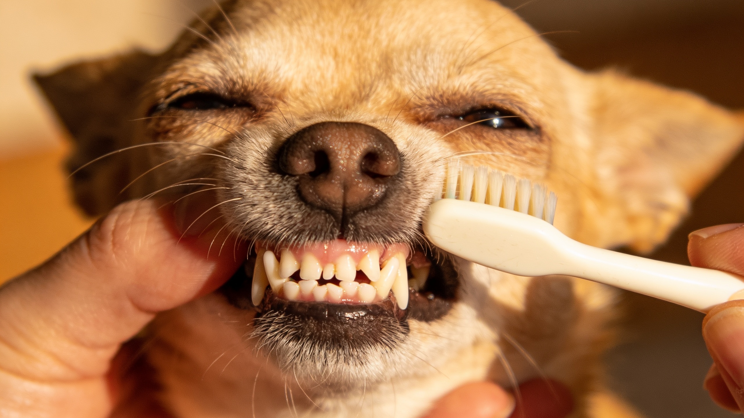
Daily tooth brushing disrupts the bacterial biofilm before it mineralizes into calculus; once plaque hardens, home care cannot remove it and the gingival inflammation continues beneath the visible tartar. Mechanical brushing is more effective than rinses, powders, or chews because the bristles shear plaque from the gingival margin, where periodontal disease begins. Use a soft canine or infant brush with a small head so the bristles can sweep along the outer surfaces of the teeth, especially the premolars and upper canines, which collect the heaviest plaque because saliva, lip conformation, and tooth shape favor retention there.
Toothpaste is mainly a carrier and flavoring agent; enzymatic formulations may help, but the decisive factor is friction and time at the gumline. Human toothpaste is unsuitable because detergents and fluoride are not meant for swallowing in dogs and can trigger gastrointestinal irritation. The most useful technique is short, calm, repeatable sessions that let the dog remain under threshold; resistance, head turning, lip tension, and tongue retraction usually indicate that pressure is too high or the handler is moving too fast. Fear responses quickly condition avoidance, so early handling of the muzzle, lips, and teeth should be paired with food reinforcement and pause-based consent.
Brachycephalic breeds such as Bulldogs, Pugs, and Boston Terriers often have crowded dentition and rotated teeth, which create plaque-retentive niches and make home cleaning more demanding. Small breeds, including Yorkshire Terriers, Dachshunds, and Chihuahuas, are overrepresented in periodontal disease because tooth crowding, retained baby teeth, and a relatively small jaw increase plaque accumulation and gingival trauma. Working breeds that mouth objects heavily may wear tooth surfaces but still develop disease at the gumline if brushing is absent. Puppies should be introduced early, before the period of greatest exploratory biting and jaw sensitivity, because habituation during this window improves long-term tolerance.
Daily inspection should include the color of the gums, the smell of the breath, and the response to gentle lip lifting. Bleeding at the brushing site is not “normal sensitivity”; it usually reflects inflamed tissue with fragile capillaries and should prompt better plaque control rather than reduced cleaning. Brown or yellow deposits along the cervical margin, drooling, unilateral chewing, dropping food, pawing at the mouth, or preference for softer foods indicate oral discomfort and possible periodontal progression. Cats may hide pain more effectively than dogs, but dogs also mask early disease by continuing to eat while chewing only on one side.

Purina Pro Plan Complete Essentials Chicken & Rice Dry Dog Food, 35 lb Bag | Shredded Blend crunchy kibble with tender pieces, real chicken as the first ingredient, live probiotics for digestive health
$74.48 (as of October 23, 2026 12:02 GMT +00:00 - More infoProduct prices and availability are accurate as of the date/time indicated and are subject to change. Any price and availability information displayed on [relevant Amazon Site(s), as applicable] at the time of purchase will apply to the purchase of this product.)High-yield home-care measures
- Brush once daily, ideally at a consistent time, because plaque matures within hours and daily disruption reduces gingival inflammation more reliably than intermittent cleaning.
- Focus on the outer tooth surfaces; the tongue naturally reduces plaque on the inner surfaces, so effort should be concentrated where it matters most.
- Use very small amounts of toothpaste to avoid swallowing excess paste and to keep the session brief and tolerable.
- Lift the lip gently rather than forcing the mouth open; force increases defensive behavior and reduces cooperative learning.
- Reward stillness and acceptance, not struggling after the brush appears; this shapes calm participation instead of panic.
- Replace worn brushes regularly, because splayed bristles lose plaque-removing efficiency at the gingival edge.
Diet can support but never replace brushing. Dry kibble alone does not reliably clean teeth because many dogs fracture kibble before meaningful abrasion occurs, and the crumbly residue can still feed plaque-forming bacteria. Dental diets with larger, fibrous kibble structures may reduce plaque by mechanical action, but they work best as adjuncts. Chews should be selected for compressibility rather than hardness; items that are hard enough to resist indentation can fracture enamel, especially in dogs with enamel defects, malocclusions, or a history of slab fractures. Controlled chewing on veterinary oral-health products can help, yet it should be viewed as oral hygiene support, not equivalent to brushing.
For dogs with severe handling sensitivity, start with systematic desensitization: touch the cheek, reward calmness, briefly lift the lip, reward, then progress to a fingertip rubbing the gumline before introducing a brush. The dog must remain relaxed enough to take treats, blink normally, and keep loose facial muscles; if arousal rises, the step was too large. This approach is especially valuable in rescue dogs, individuals with previous oral pain, and breeds predisposed to guarding when restrained. Chronic oral discomfort can sensitize even gentle dogs, so aversion to brushing should be interpreted as a possible pain signal, not stubbornness.
Periodontal disease is the most common oral disorder in dogs and begins with bacterial plaque at the gingival margin, where oxygen is limited and anaerobic organisms proliferate. The first lesion is gingivitis: reddening, swelling, and bleeding of the free gum caused by inflammatory infiltration and breakdown of the epithelial attachment. At this stage the process is still reversible if plaque is consistently disrupted, but once infection extends below the gumline and periodontal ligament fibers are destroyed, the tooth loses support and bone resorption follows. Toy breeds and dogs with crowded mouths often show marked disease despite relatively little visible tartar, because the depth of the pocket, not the surface stain, determines severity.
Retained deciduous teeth are a frequent problem in small and brachycephalic dogs because the permanent tooth erupts in an already crowded arch and cannot assume its normal position. The retained baby tooth traps food and plaque between two crowns, creating a closed, inflamed niche that accelerates gingivitis and may displace the adult tooth. Malocclusion can also injure the palate, lips, or opposing teeth; slab fractures and traumatic wear are common in dogs selected for intense gripping, tugging, or object carrying. Any tooth that seems out of line, mobile, or painful on prehension deserves prompt evaluation, because abnormal contact patterns perpetuate inflammation and secondary infection.
Fractured teeth are common in dogs that chew antlers, bones, hooves, hard nylon toys, or stones. The crown may appear intact while the pulp cavity is exposed microscopically or by a small chip, allowing oral bacteria to enter the pulp and trigger irreversible pulpitis, necrosis, and a draining periapical abscess. Enamel fractures of the carnassial teeth are especially problematic because these large cheek teeth bear high chewing forces and have complex roots; pain may show up as unilateral mastication, facial sensitivity, or refusal to play tug. Darkening of the crown after trauma can indicate devitalization even when the dog continues to eat normally.
Gingival hyperplasia, common in some genetically predisposed lines, produces redundant tissue folds that deep-cleaning at home cannot reach and that trap plaque at the sulcus. Boxers, Bulldogs, and certain giant breeds may develop exaggerated gingival overgrowth associated with chronic inflammation, hormonal influences, or breed tendency. Oral masses, including epulides and papillomas, can mimic routine dental swelling but alter airflow, prehension, and tooth cleansing by creating turbulent saliva flow and occluded spaces. Any persistent gingival lump, especially one that bleeds easily or alters the bite, should be assessed rather than assumed to be simple tartar.
Endodontic disease is often silent until the surrounding tissues react. A tooth with a nonvital pulp may develop apical infection, nasal discharge if the roots communicate with the nasal cavity, or a firm swelling under the eye when the upper fourth premolar is involved. Dogs with advanced disease may still eat because chewing instinct overrides pain, but they frequently shift to the opposite side, swallow larger pieces, or avoid hard pressure on toys. Subtle signs such as lip licking, increased salivation, reluctance to let the head be touched, or repeated yawning can reflect oral pain before obvious drooling or inappetence appears.
When one tooth changes color, position, or behavior around touch, assume a pathologic process until proven otherwise; dogs rarely “grow out of” oral discomfort.
Professional dental care begins with a complete oral examination under anesthesia, because awake inspection cannot assess the subgingival pocket, root surface, or periodontal attachment accurately. Sedation alone is usually insufficient for a thorough procedure; the patient must be immobile enough for charting, probing, radiography, scaling below the gumline, and safe management of the airway. A normal-looking crown can hide deep periodontal pockets, root exposure, or endodontic disease, so surface tartar is a poor guide to true disease burden. Full-mouth dental radiographs are not optional in a proper veterinary dental workup, since most pathology in dogs lies below the gingival margin where the eye cannot see.
Probing depth and attachment loss determine prognosis more reliably than tartar load. A tooth with shallow pockets and reversible gingivitis may be stabilised, while one with deep pockets, furcation exposure, or marked bone loss often carries a poor long-term outlook even if the crown still appears functional. Radiographs show root resorption, periapical lucency, retained roots, unerupted teeth, and jaw bone destruction, all of which alter treatment choice. They also reveal whether a painful tooth is salvageable with periodontal therapy or needs extraction to remove the nidus of chronic infection.
Scaling removes mineralized deposits above and below the gumline, but polishing is equally necessary because microscopic scratches left by instrumentation increase plaque retention if they’re not smoothed. Subgingival debridement is where the real therapy occurs; bacteria and endotoxins persist in the pocket unless the root surface is cleaned and the inflamed pocket irrigated. When attachment loss is advanced, extraction is often more humane than repeated partial cleaning, because chronic periodontal pockets remain a source of pain, halitosis, and intermittent bacteremia. Dogs with severe disease may show a rapid improvement in appetite and facial relaxation after diseased teeth are removed.
General anesthesia also allows safe treatment of fractured teeth, retained roots, oral masses, and severe gingival overgrowth. Root canal therapy can preserve strategic teeth such as the carnassials and canines when the root structure is sound and the pulp is compromised but not the surrounding support. Extraction is preferred when the tooth has severe periodontal loss, a vertical root fracture, advanced resorption, or infection extending into the surrounding bone. The choice should be based on function, root anatomy, and pain control, not on whether the crown is visible or cosmetically intact.
Findings that justify prompt veterinary oral evaluation
- Halitosis that’s foul, metallic, or worsening, because odor reflects bacterial metabolism and tissue breakdown.
- Bleeding gums, even if mild, since healthy gingiva should not bleed with routine handling.
- One-sided chewing, dropping food, or chewing unusually slowly, which often signals focal pain.
- Facial swelling, especially below the eye or along the jaw, suggesting abscessation or root infection.
- Loose teeth, broken crowns, or a tooth that has changed color after trauma.
- Discharge from the nose, oral fistulas, or sneezing with food particles, which can indicate root-to-nasal communication.
Age changes the pattern of care. Young dogs need assessment of eruption, retained deciduous teeth, and occlusion before abnormal contacts become fixed. Middle-aged dogs often develop the first clinically relevant periodontal pockets despite years of apparently normal chewing. Seniors accumulate the consequences of prior inflammation, root exposure, tooth loss, and systemic effects from chronic oral pain. Small-breed seniors, brachycephalic adults, and dogs with historical crowding should be examined more frequently because their disease progresses silently until support tissue is already lost.
Breeds selected for jaw shape or task often carry predictable dental risks. Greyhounds and other sighthounds commonly show heavy calculus and advanced periodontal disease with relatively little warning because their long, narrow skulls concentrate plaque along crowded cheek teeth. Retrieval and gripping breeds may fracture canines and carnassials during work or play, while brachycephalic dogs suffer from rotated teeth, persistent deciduous teeth, and reduced self-cleansing due to compressed arches. Knowing the breed’s functional history helps predict where pain will start and which teeth need the closest surveillance.
After treatment, oral pain control matters because untreated postoperative pain reduces food intake, increases guarding, and can make future handling more difficult. A dog that has had extractions or deep periodontal therapy may need several days of softened food, careful monitoring of appetite, and restricted access to hard chews that could disturb the healing socket. Recheck examination is used to confirm tissue closure, reassess remaining teeth, and plan the next preventive interval, because periodontal disease is managed long term, not cured by a single cleaning.
Related Posts

How to Transition Your Dog to a New Diet Safely

Calming Dog Bed Mat